Silver

Sistema de instrumentación pedicular toraco-lumbo-sacro-pélvico de titanio con recubrimiento de plata antibacteriano.

Recubrimiento de plata antibacteriano Silver

Reducción del riesgo de infección aguda y subaguda.

Implantes esterilizados individualmente.

Disminución del tiempo de hospitalización.

Compatibilidad con RX, TAC, RNM, GGO, PET y SPECT

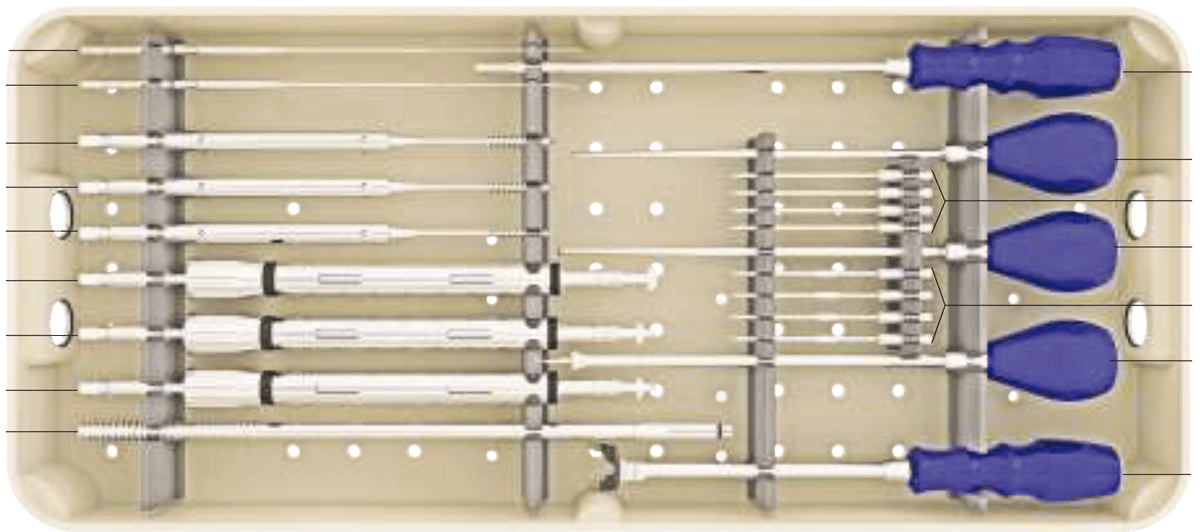
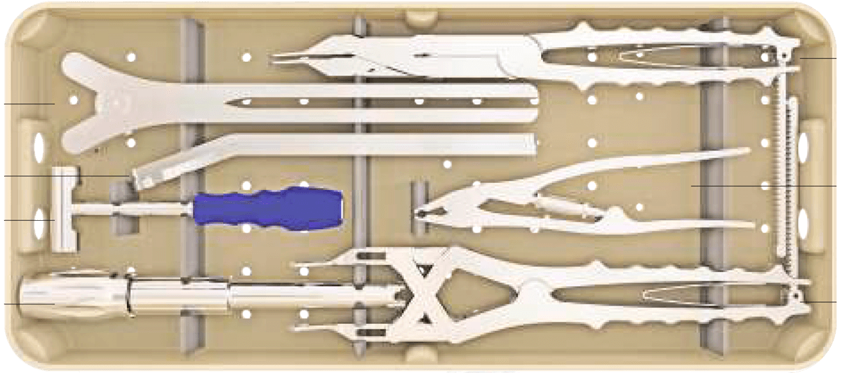
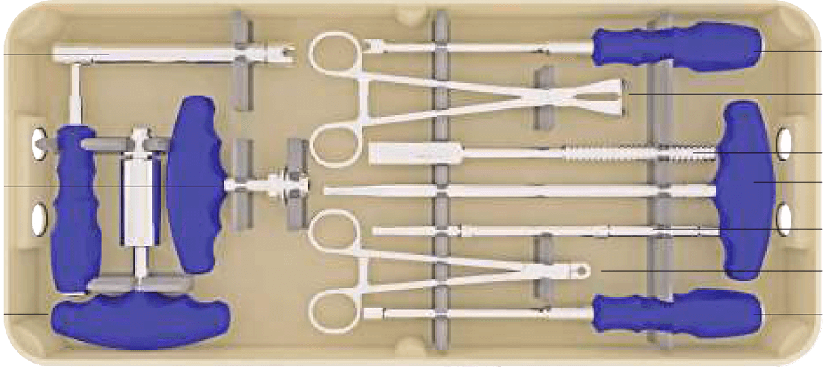
Cajas de instrumentación Silver

Tecnología única

El sistema espinal recubierto de nanopartículas de plata es una tecnología única, que por sus características antibacterianas reduce la tasa y el riesgo de infección aguda y subaguda en intervenciones de columna.

Reducción de las tasas de infección

La duración de las operaciones de estabilización o fusión de columna mediante sistema de tornillos pediculares y barras, que es uno de los procedimientos rutinarios en cirugía de columna, los factores predisponentes a la infección como las grandes incisiones, y la implementación de materiales (tornillos de aleación de titanio), conllevan un riesgo aumentado de infección.

En el caso de que la misma se produzca, las antibioterapias endovenosas y el aumento de los días de ingreso hospitalario, incrementan grandemente el coste del tratamiento. Adicionalmente, los antibióticos de que disponemos no siempre son suficientes para solucionar el problema, y en algunos casos, la retirada del material, lo que conlleva una segunda intervención, es necesario.

Esto afectará negativamente la calidad de vida del paciente, el tiempo de hospitalización, el coste del tratamiento, y la confianza del paciente tanto en el cirujano como en el centro hospitalario. La bibliografía reciente sostiene que los implantes de titanio recubiertos de nanopartículas de plata que se han implementado en cirugías de alto riesgo de infección en Traumatología y Ortopedia reducen la tasa de infección. Los centros con más experiencia en este tipo de implantes son el Royal Orthopaedic Hospital Oncology Service, Royal Orthopaedic Hospital NH Foundation Trust (Birmingham, UK), y el Department of Orthopedics and Tumor Orthopedics, Münster University Hospital (Münster, Germany), que desde 2015 publican los resultados de sus megaprotesis tumorales recubiertas de nanoparticulas de plata¹ ² ³.

Certificaciones

El Sistema Silver de instrumentación toraco-lumbo-sacro-pélvica cuenta con las certificaciones CE, ISO 9001, ISO13485, FDA y directiva de dispositivos médicos 93/42/CEE.

Los implantes de plata

Actualmente, la plata está ampliamente utilizada en el campo de los implantes de odontología y a grandes concentraciones, como cauterizador.

La plata es un material algodinámico, es decir, que permite la emisión espontánea de iones que están en su superficie, y estos iones son antibacterianos. El cobre, el zinc y el oro son también materiales algodinámicos, pero por su coste y por algunas características tóxicas, no tienen las ventajas de la plata.

Están ampliamente estudiadas las características bactericidas y bacteriostáticas de la plata⁴ ⁵ ⁶ sobre los microorganismos eucariotas (bacterias y hongos) por diferentes mecanismos: produce enzimas que evitan la mitosis, anulando la replicación del DNA bacteriano; eliminando la impermeabilidad de las membranas celulares produciendo así una insuflación del microorganismo y la consecuente apoptosis del mismo; alterando el metabolismo celular ocasionando la necrosis del germen.

Los implantes del Sistema Silver están fabricados en aleación de Titanio (Ti6AL4V) + recubrimiento con nanopartículas de plata por la ruta sol-gel a partir de nitrato de plata⁷ ⁸ ⁹. Está testado que el sistema espinal de titanio recubierto de plata mediante el método de la nanotecnología tiene un efecto antibacteriano eficaz manteniendo las características físicas y biomecánicas del material *.

El volumen de recubrimiento de plata en los implantes se realiza en un grosor de micras, y en un peso de microgramos por biocompatibilidad *. Una vez instrumentado un paciente un paciente con implantes con recubrimiento de plata, se producen unos aumentos del nivel de plata en torrente sanguíneo hasta un máximo de 5 mg. El cuerpo humano en su estado normal posee aproximadamente 1 mg de plata. La menor cantidad de plata documentada que ha producido argiria es de 4-5 gramos. Así, los niveles máximos de plata en sangre adquiridos post implementación de implantes recubiertos de nanopartículas de plata (máximo 6 mg) ni se acercan a los niveles tóxicos que causarían argiria aguda o crónica.¹⁰ ¹¹

Los productos se sirven estériles por el método de óxido de etileno. Si el producto, por cualquier motivo se desesteriliza, se puede reesterilizar en autoclave (134°C 5 min). No se deberían utilizar si existe alguna duda sobre la esterilidad del producto.

Los implantes son compatibles con RX, TAC, RNM, GGO, PET y SPECT.

Evidencia científica de implantes de titanio recubiertos de plata

Michael C. Parry, Minna K. Laitinen, Jose I. Albergo, Czar L. Gaston, Jonathan D.

Stevenson, Robert J. Grimer, Lee M. Jeys. To appear in: European Journal of Surgical Oncology. PII: S0748-7983(18)32031-6

DOI: https://doi.org/10.1016/j.ejso.2018.12.009

Reference: YEJSO 5190

Received Date: 3 August 2018/ Revised Date: 21 November 2018/ Accepted Date: 11 December 2018

Royal Orthopaedic Hospital Oncology Service, Royal Orthopaedic Hospital NH Foundation Trust, B31 2lAP Birmingham, UK

Department of Orthopaedics and Traumatology, Helsinki University General Hospital, Topeliuksenkatu 5, 00260, Helsinki, Finland

Hospital Italiano Buenos Aires, 4190 Buenos Aires, Argentina

Department of Orthopaedics, Musculoskeletal Tumor Unit, Philippine General Hospital, Manila, Philippines 1000.

Arne Streitbuerger,  Marcel P. Henrichs, Gregor Hauschild,  Markus Nottrott,  Wiebk Guder, Jendrik Hardes. European Journal of Orthopaedic Surgery & Traumatology. https://doi.org/10.1007/s00590-018-2270-3

Received: 29 April 2018 / Accepted: 14 June 2018/ Published online: 20 June 2018.

Department of Orthopedics and Tumor Orthopedics, Münster University Hospital, Münster, Germany

Department of Oncologic Musculoskeletal Surgery, Essen, University Hospital, Hufelandstr. 55, 45147 Essen, Germany

Department of Orthopedics, Paracelsus Clinic, Osnabrück,Germany

H. Wafa, R. J. Grimer, K. Reddy, L. Jeys, A. Abudu, S. R. Carter, R. M. Tillman. The Bone Joint & Joint Journal. 2015;97-B:252–7. doi:10.1302/0301-620X.97B2.34554

Received 29 May 2014/ Accepted after revision 21 October 2014.

The Royal Orthopaedic Hospital NHS Trust, Birmingham, United Kingdom

Kutsal DEVRİM SEÇİNTİ, MD, OnurÖZGÜRAL MD, Hakan TUNA MD, Ayhan ATTAR MD. Original Research.

Received: 19.05.2008

Accepted: 11.10.2008

Department of Neurosurgery, Ankara University Faculty of Medicine, Ankara

Lingzhou Zhao, Paul K. Chu, Yumei Zhang, Zhifen Wu. Wiley InterScience (www.interscience.wiley.com). DOI: 10.1002/jbm.b.31463.

Received 5 March 2009/ revised 9 May 2009/ accepted 13 May 2009/ Published online 27 July 2009

School of Stomatology, The Fourth Military Medical University, Xi’an 710072, People’s Republic of China

Department of Physics and Materials Science, City University of Hong Kong, Kowloon, Hong Kong, People’s Republic of China

Kutsal Devrim Secinti, Murat Ayten, Gokmen Kahilogullari, Gulsah Kaygusuz, Hasan Caglar Ugur, Ayhan Attar. Journal of Clinical Neuroscience 15 (2008) 434–439.

Received 5 September 2006/ accepted 7 March 2007

Department of Neurosurgery, Ankara University School of Medicine, Ankara, Turkey

Department of Pathology, Ankara University School of Medicine, Ankara, Turkey

Kutsal Devrim Secinti, Hakan Ozalpa, Ayhan Attara, Mustafa F. Sargon. Journal of Clinical Neuroscience 18 (2011) 391–395.

Received 21 February 2010/ Accepted 14 June 2010

Department of Neurosurgery, School of Medicine, University of Ankara, Samanpazari, Sihhiye, Ankara 06100, Turkey

Department of Anatomy, School of Medicine, University of Hacettepe, Sihhiye, Ankara, Turkey

Ziqiang Xu, Man Li, Xia Li, Xiangmei Liu, Fei Ma, Shuilin Wu,  K. W. K. Yeung, Yong Han and Paul K. Chu. ACS Applied Materials & Interfaces. Research Article. DOI: 10.1021/acsami.6b04161

Received: April 7, 2016/ Accepted: June 14, 2016

State Key Laboratory for Mechanical Behavior of Materials, School of Materials Science and Engineering, Xi’an Jiaotong University, Xi’an, Shaanxi, 710049, China

Hubei Collaborative Innovation Center for Advanced Organic Chemical Materials, Ministry-of-Education Key Laboratory for the Green Preparation and Application of Functional Materials, Hubei Key Laboratory of Polymer Materials, School of Materials Science & Engineering, Hubei University, Wuhan, 430062, China

Division of Spine Surgery, Department of Orthopaedics & Traumatology, Li KaShing Faculty of Medicine, The University of Hong Kong, Hong Kong, 999077, China

Department of Physics & Materials Science, City University of Hong Kong, Tat Chee Avenue, Kowloon, Hong Kong, 999077, China

Derya Burcu Hazer, MD, Mustafa Sakar, MD, Yelda Dere MD, Gu¨ ls¸en Altınkanat, MD,  M. Ibrahim Ziyal, MD, and Baki Hazer, PhD. Spine Volume 41, Number 6, pp E323–E329_ 2016. DOI: 10.1097/BRS.0000000000001223

Acknowledgment date: June 2, 2015. First revision date: July 15, 2015.

Second revision date: August 11, 2015. Acceptance date: September 7,

2015.

Mug˘la SıtkıKoc¸man University, Faculty of Medicine, Department of Neurosurgery, Mug˘la, Turkey

Marmara University, Institute of Neurological Sciences, Istanbul, Turkey; zMug˘la SıtkıKoc¸man University, Faculty

of Medicine, Department of Pathology, Mug˘la, Turkey

Marmara University, Faculty of Medicine, Department of Microbiology, Istanbul, Turkey

Marmara University, Faculty of Medicine, Department of Neurosurgery, Institute of Neurological Sciences, Istanbul, Turkey

Bu¨ lent Ecevit University, Department of Chemistry, Engineering of Nanotechnology, Zonguldak, Turkey.

B. Hussmann, I. Johann,1 M. D. Kauther, S. Landgraeber, Marcus Jäger and S. Lendemans. BioMed Research International. Volume 2013, Article ID 763096, 11 pages. http://dx.doi.org/10.1155/2013/763096

Trauma Surgery Department, University Hospital Essen, Hufelandstraße 55, 45122 Essen, Germany

Orthopaedic Department, University of Duisburg-Essen, Hufelandstraße 55, 45122 Essen, Germany

Guido Scoccianti, MD, Filippo Frenos, MD, Giovanni Beltrami, MD, Domenico Andrea Campanacci, MD, Rodolfo Capanna, MD. Injury. http://dx.doi.org/10.1016/j.injury.2016.07.042 0020-1383/ã 2016 Elsevier Ltd. All rights reserved.  Orthopaedic Oncology Unit, Careggi University-Hospital, Firenze, Italy

Silver has been used to reduce infection for centuries. This study retrospectively analyzed whether the introduction of silver-impregnated dressing (SD; Silverlon, Argentum Medical, LLC, Lakefront, GA) rather than RD (iodine- or alcohol-based swab and dry 4 × 4 gauze) would reduce the risk of superficial or deep infection after lumbar laminectomy with instrumented fusion.

Infection in spinal implant is of great concern. Anti-infection strategies must be tested in relevant animal models that will lead to appropriate clinical studies.

Surgical site infection (SSI) after hip fracture surgery is a well-known complication with serious consequences for both the patient and the medical system. Silver ion treatment is considered an effective antibacterial agent, however, the use of silver dressing (SD) in the primary prevention of SSIs is controversial. The aims of this study were to compare SD with regular dressing (RD) in the prevention of SSI in elderly patients undergoing surgery for hip fractures, and to compare costs.

Pin tract infection is a frequent complication of external fixation; according to literature its frequency ranges from 2–30%. The recent introduction of silver coating of polymeric materials was found to decrease bacterial adhesion; its clinical use with Foley catheters and central venous catheters led to significant results. To verify the ability of the same silver coating to decrease the bacterial colonization on external fixation screws, a prospective randomized study was carried out on 24 male patients; a total of 106 screws were implanted in the lower limb to fix femoral or tibial diaphyseal fractures: 50 were coated with silver and 56 were commercially available stainless steel screws. Although the coated screws resulted in a lower rate of positive cultures (30.0%) than the uncoated screws (42.9%), this difference was not statistically significant (p 5 0.243). the clinical behavior of the coated screws
did not differ from that of the uncoated ones. Furthermore, the implant of silver-coated screws resulted in a significant increase in the silver serum level. These results led us to consider it ethically unacceptable to continue this investigation.

In this study, electrolytically deposited strongly adherent silver nanoparticles on stainless-steel (SS) implants were used for in situ osteomyelitis treatment. Samples were heat treated to enhance adhesion of silver on 316 L SS. Ex vivo studies were performed to measure silver-release profiles from the 316 L SS screws inserted in equine cadaver bones. No change in the release profiles of silver ions were observed in vitro between the implanted screws and the control. In vivo studies were performed using osteomyelitic rabbit model with 3 mm diameter silver-deposited 316 L SS pins at two different doses of silver: high and low. Infection control ability of the pins for treating osteomyelitis in a rabbit model was measured using bacteriologic, radiographic, histological, and scanning electron microscopic studies. Silver-coated pins, especially high dose, offered a promising result to treat infection in animal osteomyelitis model without any toxicity to major organs.

Bio-implants in the human body act as passive surfaces that are prone to bacterial adhesion potentially leading to deep body infections. Pedicle screws made of uncoated or silver-coated titanium alloy were used both in vitro and in vivo to determine whether silvercoated materials have antimicrobial properties when they are anodized. Twenty-four New Zealand Albino rabbits were divided into four groups with six in each. In Group 1, the rabbits were exposed to 8 lA direct current (DC) via silver-coated screws. In Group 2, the rabbits were not exposed to any electrical current, but silver-coated screws were used. In Group 3, the rabbits were exposed to 8 lA DC using uncoated screws. In Group 4, the rabbits were not exposed to any electrical current, but uncoated screws were used. Staphylococcus aureus (106 cfu) was inoculated into the rabbits before any electrical current was applied. All the animals were killed, and the areas surrounding the screws were histologically and microbiologically examined. Silver-coated titanium screws prevented implant-associated deep bone infections when they were polarized anodically. The antibacterial effects of the same screws with the same bacterium were confirmed in in vitro experiments on agar plates. When the screws were anodized with the same electrical parameters in vitro, a marked inhibition zone was detected around the silver-coated screws but not around the uncoated screws. Our findings suggest that silver-coated titanium implants can be used to prevent implant-associated deep bone infections when they are polarized anodically.

The formation of bacterial biofilm on the surface of implanted metal objects is a major clinical problem. The antibacterial and antifungal effect of silver ions has been long known, and seems to give silver the capability to inhibit biofilm formation. To test the effect of silver ions, 20 New Zealand rabbits had bacteria applied to a screw insertion site at the iliac crest, and were then randomly divided into two groups: Group I, which had silver-coated screws applied, and Group II, which had uncoated titanium screws. After the rabbits were sacrificed on day 28, we examined the screws, the bone adjacent to the screws, and the liver, kidneys, brain and corneas of both groups under transmission (TEM) and scanning electron microscopy (SEM). We also analysed microbiological samples from the screw holes. All silver-coated screws, but only 10% of uncoated titanium screws, were sterile. All tissue samples appeared ultrastructurally normal in both groups. Biofilm formation was inhibited on all silver-coated screws, but all uncoated screws developed a biofilm on their surfaces. Our findings suggest that nanoparticle silver ion-coated implants are as safe as uncoated titanium screws and that they can help prevent both biofilm formation and infection.

Otros artículos:

Implant-related infection of biomaterials is one of the main causes of arthroplasty and osteosynthesis failure. Bacteria, such as the rapidly-emerging Multi Drug Resistant (MDR) pathogen Acinetobacter Baumannii, initiate the infection by adhering to biomaterials and forming a biofilm.

Antibacterial coatings (ABCs) of implants have proven safe and effective to reduce post-surgical infection, but little is known about their possible economic impact on large-scale use. This study evaluated the point of economic balance, during the first year after surgery, and the potential overall annual healthcare cost savings of three different antibacterial technologies applied to joint arthroplasty: a dual antibiotic-loaded bone cement (COPAL G+C®), an antibacterial hydrogel coating (DAC®) and a silver coating (Agluna®).

Nanometer scale surface features on implants and prostheses can potentially be used to enhance osseointegration and may also add further
functionalities, such as infection resistance, to the implant. In this study, a nanostructured noble metal coating consisting of palladium, gold
and silver, never previously used in bone applications, was applied to machined titanium screws to evaluate osseointegration after 6 and
12 weeks in rabbit tibiae and femurs.

Retrospective studies of silver-coated hip implants have demonstrated promising results and safety profile, however, the potential benefits are so far unproven in prospective studies. Silver-coated implants may have a role in patients undergoing revision or primary surgery with a high risk of infection but as yet there are no human studies investigating silver in primary hip arthroplasty. Adequately powered robust prospective studies are needed in this area to determine if silver-coated implants would be efficacious and cost-effective. The purpose of this systematic review article is to review the current literature regarding the use of silver in hip arthroplasty. Our review showed that there is some encouraging evidence that silver coatings can reduce infection.

Implant-associated infections remain a major issue in orthopaedics and antimicrobial functionalization of the implant surface by antibiotics or other anti-infective agents have gained interest. The goal of this article is to identify antimicrobial coatings, for which clinical data are available and to review their clinical need, safety profile, and their efficacy to reduce infection rates.

*SILVER Biomechanical Data & Biocompatibility Report

Visita nuestra galería de fotos